

FREE Complications Webinar
Want to overcome your fear of complications and confidently master anatomy?
Join us for one of Dr Tim's FREE upcoming webinars.
Check dates here and save your spot
 Dr Tim Pearce
One of the worst vascular occlusion cases doesn’t begin with the injection, it begins with what happens after. A patient rang her clinic within three hours, worried something was wrong. The clinic reassured her that pain after filler was normal. She called back later with the same concerns, receiving the same reassurance. When blisters appeared, the clinician diagnosed cold sores. Eventually, in severe pain, she went to A&E where staff treated her for a skin infection.
The entire time, the window to dissolve the filler blocking her blood supply was closing. One diagnostic mistake led to extensive necrosis, entirely preventable with systematic thinking about diagnosis timing.
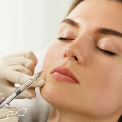
A particularly painful injection has been the first reported sign in many vascular occlusion cases. This creates a diagnostic challenge because every injection causes some discomfort. You need to assess whether this specific injection feels disproportionately uncomfortable compared to others in the same treatment session.
The test isn’t highly sensitive, but clinicians often recognize in retrospect that the injection felt distinctly different. When an injection hurts more than others on the same patient, stop immediately. Withdraw the needle, check capillary refill, and confirm the patient isn’t hemorrhaging from the injection site (which signals you’ve penetrated a vessel). Choose a different position for the next injection attempt.
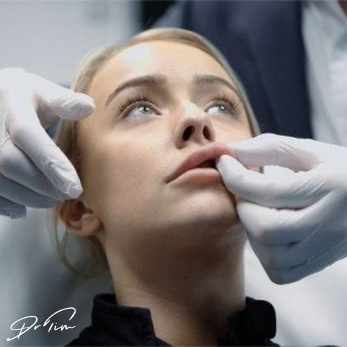
Pallor typically appears as the second warning sign. A true complete vascular occlusion produces stark blanching, especially visible in lips, a gray patch with zero capillary refill. These complete occlusions are obvious in lip tissue but easier to miss on other facial areas or in patients with darker skin tones.
False positives complicate diagnosis. Patients develop pallor without vascular occlusion after using lidocaine anesthetic cream, which creates blotchy discoloration on lips. The procedure itself can cause similar blotchiness, confusing both patients and injectors.
The critical distinction appears on close examination: temporary blanching from anesthetic shows capillary refill within two seconds. The skin looks paler, but blood returns. Compress the lip and check, if you see any capillary refill in the pale area’s center, you’re seeing anesthetic-induced blanching.
Anatomical distribution provides another clue. When both upper and lower lips show blotchiness, you’ve probably not occluded both superior and inferior labial arteries. The lidocaine effect explains this pattern better than simultaneous dual arterial occlusions.
Pallor doesn’t persist long. As an early sign, it typically transitions to the next stage within minutes.
This net-like pattern, blotchy purple rash, indicates disrupted blood drainage. Venules may still remove blood from skin tissue, but arteries fail to replenish it. Small pale patches alternate with darker areas, creating the characteristic mottled appearance associated with larger vascular occlusions.

Livedo reticularis develops over minutes, not seconds. The blotchiness typically starts 15 to 30 minutes after injection and requires immediate clinical assessment.
Post-procedure discomfort is normal, most patients report achiness or tenderness after substantial treatment in multiple areas. The diagnostic challenge lies in distinguishing normal discomfort from vascular occlusion pain.
Look for localized pain significantly worse than other treated areas. When patients request strong painkillers to manage their discomfort, arrange immediate evaluation.
Multiple cases involve patients who didn’t know pain signaled a potential complication. They assumed post-procedure pain was normal, took painkillers, went to sleep, and woke hours later with progressed vascular injury. One severe nose occlusion case involved a patient who took Tramadol for what she assumed was normal pain, slept through the night, and woke to find advanced tissue damage.
Teach patients to distinguish discomfort from pain. Tenderness when touching treated areas or resting your head on a pillow qualifies as normal discomfort. Building, throbbing pain concentrated in one location requires evaluation.
Blister diagnosis causes frequent confusion in aesthetics practice. One infamous case involved a salon owner who accused a patient with perioral blisters of having excessive oral sex, a diagnostic error that represents common clinician mistakes with this complication.
Herpes causes pale blisters. Necrotic tissue breakdown produces pustules, yellowy blisters that follow arterial pathways. Necrosis-related blisters rarely affect the white lip, appearing instead on the pink lip tissue.

When patients report post-procedure blisters, verify the location. Request photographs. Confirm the presentation matches that specific patient’s herpes history rather than representing a new event with different blister characteristics. New yellowy blisters in arterial distribution patterns indicate early tissue breakdown.
Check capillary refill on all patients throughout your injection session, not just at the end. While cleaning skin and checking for bleeding, verify that capillary refill returns quickly. This simple assessment catches complications faster than any other diagnostic method.
One clinic tracked 17 vascular occlusions across eight clinicians over 15 years. Every single case was identified immediately on the treatment day, treated comprehensively, and resulted in zero necrotic wounds. Consistent capillary refill checking made this perfect record possible.
Completing a career without causing necrotic injury remains achievable. You’ll likely encounter vascular occlusions, that’s different from causing tissue necrosis. Actual tissue breakdown becomes avoidable when you address each risk factor systematically.
Reduce the probability of intra-arterial injection through anatomical knowledge, ultrasound use, and aspiration technique. These pre-injection protocols decrease the chance of needle bevel placement in vessels.
Diagnose vascular occlusions before patients leave your clinic. Check every patient’s capillary refill and educate them about warning signs.
Triage aggressively when patients report concerning symptoms. Get them back to your clinic immediately rather than waiting hours or days.
Diagnose comprehensively during acute management. Vascular occlusions affect multiple areas simultaneously.
In urgent situations, injectors often hyperfocus on one pallor area and miss secondary occlusions inside the mouth or nose that remain untreated. Consider which facial structures the occluded artery supplies and check all dependent tissue.
Maintain emergency protocols you can access instantly. You probably haven’t managed a vascular occlusion in years, this pattern characterizes most well-run clinics. Stock sufficient hyaluronidase to treat standard occlusions through the first critical hours. Know the location of your nearest hyperbaric oxygen facility and ultrasound access points.
Many practitioners avoid discussing vascular occlusion with patients, believing that risk disclosure reduces treatment uptake. This assumption reverses the actual dynamic. Patients seek practitioners they trust.
When you convey confidence describing vascular occlusion, explaining your prevention strategies and emergency protocols, you demonstrate the capability that attracts patients. Clinicians who transparently discuss complication management attract more clients than practitioners who avoid these conversations, because patients recognize genuine expertise and preparation.
Educate patients about vascular occlusion in ways that demonstrate your competence rather than creating fear. Patients positioned with a competent, prepared practitioner feel safer than patients who sense their injector avoids discussing complications.
Join us for one of Dr Tim's FREE upcoming webinars.
Check dates here and save your spot

Dr Tim Pearce MBChB BSc (Hons) MRCGP founded his eLearning concept in 2016 in order to provide readily accessible BOTOX® and dermal filler online courses for fellow Medical Aesthetics practitioners. His objective was to raise standards within the industry – a principle which remains just as relevant today.
Our exclusive video-led courses are designed to build confidence, knowledge and technique at every stage, working from foundation level to advanced treatments and management of complications.
Thousands of delegates have benefited from the courses and we’re highly rated on Trustpilot. For more information or to discuss which course is right for you, please get in touch with our friendly team.