You may be interested


 Dr Tim Pearce

When it comes to learning about facial anatomy, aesthetic clinicians and trainers usually reach for a textbook, but what if the textbooks are wrong? What do we do when what we think we ‘know’ about anatomy is wrong, and the patient in front of us has ‘weird anatomy’ or anatomical anomalies?
On 23rd November 2021, Dr Tim Pearce hosted a Live on Instagram and interviewed renowned surgeon and anatomist Dr Patricia Oyole to discuss how aesthetic clinicians can inject safely, despite ‘weird anatomy’.
In this blog, Dr Tim Pearce highlights a handful of the stand-out points from his lengthy discussion with Patricia and invites you to catch up with the whole interview now hosted on You Tube as part of the Aesthetic Mastery Show. They touch on treating the forehead, the temples, the nose, and discuss concepts such as compression necrosis and the impact of bone structure on non-surgical rhinoplasty.
Dr Tim will be discussing more medical aesthetic training tips as part of his upcoming webinar series, so if you’re looking to increase your CPD-certified learning and want to learn more skills to make you a better clinician, then step one is to register for the free webinars by Dr Tim.
Understanding veins and arteries
Kicking off the debate, Dr Patricia and Dr Tim agreed that the location and size of facial arteries, and their functional status within the treatment area are never the same in each patient. The aesthetic clinician must therefore develop a working model of what they think they know about anatomy but be prepared for surprises!
Dr Patricia noted that the trick is to learn to deal with finding structures where you are not expecting them to be located. She explained that in academia, when teaching and documenting anatomy, they present the averages, the common consensus based on anatomical studies and dissections, but this can have its flaws, due to cadaver sample sizes and extreme differences which influence the averages. As clinicians, you only have control over the technique you use when injecting and the material or product you are using, you cannot control the anatomy. You must therefore have a respectful approach towards the anatomy of the individual patient you are about to treat and deal with what you find.
Here are some of the further discussion points and questions asked by Dr Tim during his interview with Dr Patricia.
What is the best approach to treating the forehead?
 Dr Patricia said that it starts with feeling confident in your treatment plan, which also helps to put the patient at ease. She asks her patients to bend forward to bend their head over; this helps with the visibility of their vasculature.
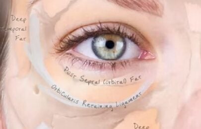
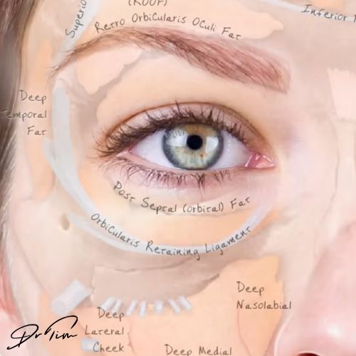
She explained how much she likes Dr Tim’s limited edition facial anatomy posters which are available to buy for your aesthetic business as a helpful tool to understanding the paths of the facial vasculature. Dr Patricia noted how the posters show the arteries and veins, including how the venous vasculature sits slightly behind the arterial vasculature
The veins in the face are a greenish colour under the skin, compared to the arteries, the latter also having a pulse. Dr Patricia explained that by asking the patient to bend forward you can often see the arteries pulsing in their forehead, and thus are able to judge the proximal location of the veins. If you intend to inject large volumes into the forehead area, she cautions that it is important to locate these pulsations, for avoidance sake. Similarly, if you cannot see the pulsation, you can often locate a visible greenish vein and feel adjacent to it for the pulsating artery to help you locate all the danger zones.
What is compression necrosis?
Dr Patricia said that she has a personal preference for the use of cannula when performing contouring treatments but warned that a 27G cannula can be just as likely to puncture or compress a vessel as a sharp needle and encouraged practitioners to be mindful of the location and direction of the aperture of their cannula.
With her experience drawn from multiple dissections, she suggested that cannulating small vessels is actually very difficult and thus most of the vascular complications seen and reported in facial aesthetic practice are compression, rather than occlusion, with stenosis or strangulation of the vessels from the outside due to deposition of filler product within the tissue layers.
Dr Patricia highlighted that this was also evident when we see reports of partial necrosis and not full necrosis. This is due to the product placed between the fat and muscle layers compressing arteries, without a vascular occlusion from an internal clot of product material. Thus collateral anastomosis of vasculature and the partial blood flow within the compressed vessel is enough to partially sustain tissue, however a complication is still evident, as compression necrosis, and tissue is not being perfused correctly. A compressed artery will show a slow capillary refill, whereas an occluded vessel has no capillary refill at all.
What is the best approach to treating the temples?
Dr Tim asked Dr Patricia which she felt was the best approach for treating the temples – to use a cannula more superficially or periosteal injections, which Dr Tim is more familiar with performing himself. She noted that both approaches are safe if the indication is correct.
For the periosteal injection technique, under the muscle, this is best for patients who do not have a strong profile. The temporalis muscle is usually a very strong muscles so if you inject filler beneath it, but the patient has strong muscle movement then the product will be subject to repeated muscle movement and cannot be guaranteed to remain in place or have longevity.
She explained that above the SMAS there is consistent hollow space that can be filled, the ligaments or temporal septa however separates into distinct areas which can be noted when running a cannula down the tissue whereby it grips as is travels down between sections. She explained that depositing a single bolus in this area and massaging to spread is not realistic in relation to the anatomy as the divided septa is not one large hollow area, but has an aponeurotic display, or fan-like branches.
What is the best approach to treating noses?

As part of this debate on treating noses, Dr Patricia drew attention to a comparison between the skulls of three types of individuals from different descent – Caucasian, Asian and African. She noted that when aesthetic clinicians are attempting to build the nasal bridge on someone from Asian descent, for example, they should be mindful of the size of the nasal bone compared to that of someone from African descent, as clearly visible in the above image. With the Asian nose, there is a lack of bone support for building filler at the bridge or nasal root when compared to the African model. She cautioned that when performing non-surgical rhinoplasty procedures and others that aesthetic clinicians need to be mindful of the bone structure and topography of the skull which will support their soft tissue implants.
Dr Tim explained that he believes that cannulas are more likely to enter a vessel when treating the nose, plus reported complications in relation to serious vascular occlusions that affected the ocular blood supply showed an over representation of incidents with cannula use. This could be because there is limited space in the nose, the route to injection is parallel with many vessels, and your need to push quite hard to traverse through the area.
Dr Patricia stated that she believes we simply have statistically more vascular accidents reported with cannulas than with needles because when treating the bridge of the nose, clinicians perform more injections using cannulas than needles. Although agreeing with her, Dr Tim pointed out that he believes that the severity of the reported incidents is more pronounced with cannula use due to larger volume deposits. Dr Patricia agreed but concludes that there is reported data that ‘touch-up’ treatments are also responsible for blocking vasculature; these are performed within the designated healing period due to patients who are anxious for results. In this case, there is a fight for space between oedema and the originally injected material during the healing time, which is then compounded by the addition of more filler material, leading to vascular accidents that do not reflect either the initial or combined volume injected as the main cause. This is more commonly noted when treating younger patients with younger anatomy where space between tissue layers is more limited, fatty tissue is more abundant, and capillary structures are more defined. Dr Patricia warned the audience against performing ‘touch-ups’ too soon.
She finished by noting that with the reduction in fat, vasculature, and collagen matrices due to changes in the ageing anatomy, it does lend itself to the creation of more space between tissue layers for the products that we seek to place into the face for rejuvenation, making this a safer patient cohort for aesthetic treatments.
You can follow Dr Patricia Oyole on Instagram for more insight on facial anatomy.
Why not also check out these blogs for additional reading:
- 5 critical mistakes clinicians make with vascular occlusions
- 8 types of vascular injury that can cause necrosis
- Plus you can download our guide to the 13 most dangerous areas to inject which includes a map of the facial vessels
Aesthetics Mastery Show
This blog accompanies a recent Aesthetics Mastery Show, where Dr Tim Pearce and Patricia Oyole discussed how to inject safely in spite of anatomical anomalies.
They discuss patient assessment tips, compression necrosis, nose anatomy, granulomas, the danger of filler touch ups, and how the patient’s age affects their risk of vascular compromise.
Watch the full Aesthetic Mastery Show episode here:
Are you still anxious about delivering cosmetic injectables safely?
If you want to learn more about mastering medical aesthetic treatments and complications or conquering the anxiety of where to place your needle, then register for the next Dr Tim webinar.
Subscribe to our YouTube channel for really useful regular tips and advice. ![]()
Dr Tim Pearce eLearning
Dr Tim Pearce MBChB BSc (Hons) MRCGP founded his eLearning concept in 2016 in order to provide readily accessible BOTOX® and dermal filler online courses for fellow Medical Aesthetics practitioners. His objective was to raise standards within the industry – a principle which remains just as relevant today.
Our exclusive video-led courses are designed to build confidence, knowledge and technique at every stage, working from foundation level to advanced treatments and management of complications.
Thousands of delegates have benefited from the courses and we’re highly rated on Trustpilot. For more information or to discuss which course is right for you, please get in touch with our friendly team.