You may be interested


 Dr Tim Pearce
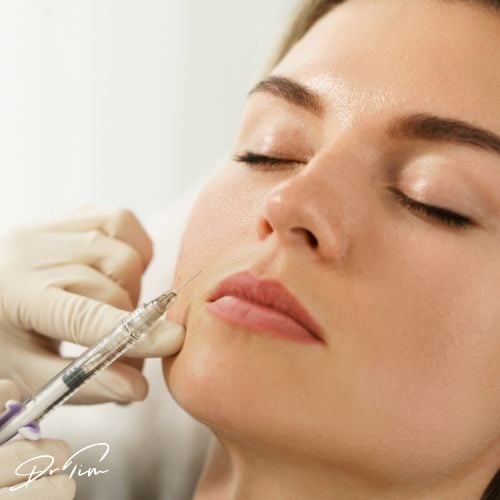
 When using dermal fillers in the lips, it is vital to understand the location of the superior and inferior labial arteries to mitigate the risk of causing a vascular occlusion and compromising blood supply to the lips and surrounding structures. But could you accurately locate them?
Sounds simple enough, and many aesthetic clinicians invest in textbooks which go to great lengths to draw diagrams and illustrations of all the vascular anastomoses within an anatomical region, but can we rely upon them in their 2D form? Published lip anatomy studies can also be contradictory, each postulating differing locations for the common position of the core arterial routes. No wonder it is such a tricky place to inject.
According to Dr Tim Pearce’s many years of personal experience, supporting and training other healthcare practitioners, the lips are the most common location on the face for causing a complication. In 2018, the ACE Aesthetics Complications Expert Group World also agreed – when auditing their private Facebook group where members go to seek peer support and advice, they found that over half of all requests for help related to lip filler treatment complications.
In this blog, Dr Tim Pearce will discuss the three-dimensional positions of the labial arteries and review recent published studies on lip anatomy.
Dr Tim will be discussing more medical aesthetic training tips as part of his upcoming webinar series, so if you’re looking to increase your CPD-certified learning and want to learn more skills to make you a better practitioner, then step one is to register for the free webinars by Dr Tim.
How to reduce your risk when injecting lip fillers
In a study entitled ‘rates of vascular occlusion associated with using needles vs cannulas for filler injections’ published by Dr Murad Alam et al, in the Journal of American Medical Association (JAMA) Dermatology, they studied the risk of injury in a cohort of dermatologist injectors and found a rate of occlusion of 1 in every 6,000mls of filler product delivered in lip treatments. More interestingly, they showed a decrease in risk as the injector gained more experience in delivering the treatments, an observation that was clearly visible, concluding that after five years, an injector has 70% less chance of causing a vascular occlusion.
This is great news, especially for those of you who are still in the early stages of your journey into aesthetic practice, because it shows that ‘practice’ – and understanding the nuances of your injection technique to mitigate risk – really does make ‘perfect’, (well almost, as we know, vascular occlusions can still happen, even in the most experienced hands). Put bluntly, we can all get safer.
What differentiates an experienced filler injector from an inexperienced injector?
Dr Tim believes that this starts with anatomical understanding – honing that three-dimensional, high-resolution image in your mind of anatomical structures, positioning, and interactions. An understanding of where the arteries are located will drive the cautious injector to be more precise when injecting.
Anyone can claim to know the anatomy by naming the arteries, but many practitioners will have a much lower resolution image in their head of the underlying anatomy, despite knowing all the right words and looking at pencil drawings.
He believes it is the time spent understanding the subtle detail of the anatomy and then using that to inform injection technique that will cut risk down in the longer term.
Dr Tim advises all aesthetic clinicians to really spend time learning and honing your skills on your mental image of the anatomy so that it is front and centre when you look at every patient sat in front of you.
Lip anatomy and labial arteries
Dr Tim has highlighted some great references made by Prof. Sebastian Cotofana, firstly looking at his published clinical paper, entitled ‘distribution pattern of the superior and inferior labial arteries: impact for safe upper and lower lip augmentation procedures’.
The paper explains not only the common depths of the relative positions of the arteries, but also the likelihood of them changing planes as they traverse the lips.
This highlights the need for all aesthetic clinicians to understand anatomy in terms of layers, placing structures with depth, so we can build up that 3D image in our minds.
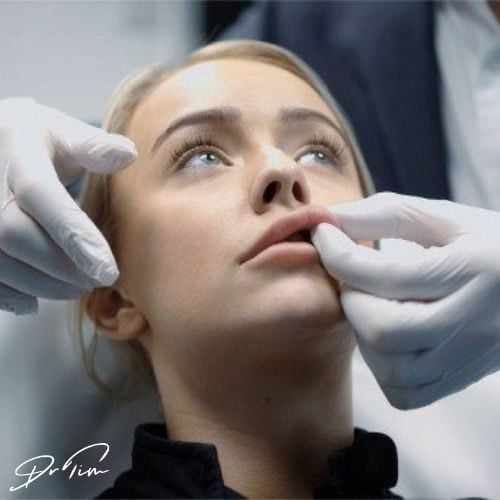
Starting from the top of the lip, we have the dermis, then a layer of hypodermic fat, then comes the orbicularis oris muscle, followed by the submucosa, and finally the mucosal layer, forming the lip envelope. The muscle inserts into the vermillion border where the divide between the mucosa and the dermis lies, and this is a very useful landmark to reference the likely position of the artery, because it is so clear to see when you are injecting.
Within Prof. Cotofana’s work, he studied 193 cadavers, a considerable sample size, and dissected at three different points along the top and again along the bottom of the lip. Findings showed that there are three layers within which one might encounter the artery – sandwiched between the oral mucosa and the muscle, within the muscle, or above the muscle in the hypodermis. 78% were posterior to the muscle, 17% were intramuscular, and 2% were in the hypodermis. Approximately 30% of the specimens showed the vessels crossing into different layers within the same cadaver. It also reported that the superior labial artery tends to run exactly where aesthetic clinicians inject dermal fillers using a variety of techniques – at the level of the vermillion border. The inferior labial artery was found to be inferior to the vermillion border, which is more helpful for injectors. However, there were approximately 2% of cases where the same lips had arteries in more than one plane, usually the subcutaneous and the submucosal.
What the findings of this paper do is reinforce the 3D notion of your anatomical understanding, 2D just will not cut it when you are injecting. Time to upgrade your mind maps to the latest imagery and get thinking in three dimensions with layers and plane of tissue as you consider placing your needle.
How reliable are cadaver studies when it comes to lips?
When approaching the lips, we know that the arteries run from lateral to medial along both the superior and the inferior labial arteries. When it comes to the depth of the arteries, multiple studies have shown that the artery usually resides beneath the orbicularis oris muscle, and the depth ranges from three to seven millimetres, depending on the paper.
However, the greatest limitation of cadaver studies is that they do not represent the normal aesthetic patient cohort of younger females, but instead are much older lips which are lacking in natural volume due to ageing. Dr Tim believes that the depth of the artery is therefore likely to become significantly different as we lose volume in the lips and this could account for discrepancies in published data, especially when comparing cadaver studies with ultrasound studies invivo.
Using ultrasound to understand lip anatomy
Prof. Cotofana also undertook an ultrasound study of a much younger cohort, with 41 volunteers with an average aged of 26, and found the average depth of the artery to be 5.6mm from the surface of the skin. The published study entitled ‘anatomy of the superior and inferior labial arteries revised: an ultrasound investigation and implication for lip volumization’ also found a different spread of arterial positioning relative to the muscle. Approximately 58% showed the artery within the submucosal plane, 36% in the muscle, and 5% in the dermis. By comparison with the cadaver study, this showed fewer below the muscle and more within, but the pattern of variation is very similar.
The most significant difference between the two papers, is that the ultrasound study describes the artery as running in the red lip and not the vermillion border, as noted in the cadaver publication.
Dr Tim believes that he has the answer – which came to him whilst cutting a tomato in half for his lunch one day!


It could be that in the ultrasound study of younger patients, the individuals had more volume in their lips and a greater outward curl of the orbicularis oris muscle. The artery in this case is deep, but as the lip ages, highlighted in the cadaver study, the muscle insertion in the vermillion falls relative to the artery which also becomes more superficial as volume is lost and so is more likely to be parallel with the vermillion border in an older patient, but slightly inferior to the border, and deeper in younger patients.
The artery is rarely IN the vermillion border, being located deeper than the border itself, but from one perspective, it is parallel with the border, and this can cause confusion. Understanding depth perception in relation to these structures is the key to mitigating the risk of vascular occlusion.
You can follow Professor Sebastian Cotofana on Instagram for more of his insight on facial anatomy.
Aesthetics Mastery Show
Dr Tim Pearce discussed the seemingly contradictory studies about the most common position of the superior and inferior labial arteries on the Aesthetic Mastery Show.
Further resources
Browse our FREE downloadable resources and access FREE eLearning by following Dr Tim on social media.
Are you still anxious about delivering cosmetic injectables safely?
If you want to learn more about mastering medical aesthetic treatments and complications or conquering the anxiety of where to place your needle, then register for the next Dr Tim webinar.
Subscribe to our YouTube channel for really useful regular tips and advice. ![]()
Dr Tim Pearce eLearning
Dr Tim Pearce MBChB BSc (Hons) MRCGP founded his eLearning concept in 2016 in order to provide readily accessible BOTOX® and dermal filler online courses for fellow Medical Aesthetics practitioners. His objective was to raise standards within the industry – a principle which remains just as relevant today.
Our exclusive video-led courses are designed to build confidence, knowledge and technique at every stage, working from foundation level to advanced treatments and management of complications.
Thousands of delegates have benefited from the courses and we’re highly rated on Trustpilot. For more information or to discuss which course is right for you, please get in touch with our friendly team.